How can we decode the cellular and molecular architecture of the brain and other organs? How can we measure and integrate different modalities of information in the same specimen? How can we relate the function of cells, tissues, and organs to their structure and molecular characteristics?
The central aim of the Danzl lab, an interdisciplinary team of neuroscientists, biologists, computer scientists, and physicists, is to develop technologies for extracting new types of information from biological specimens and address problems of biological and biomedical relevance in unprecedented ways.
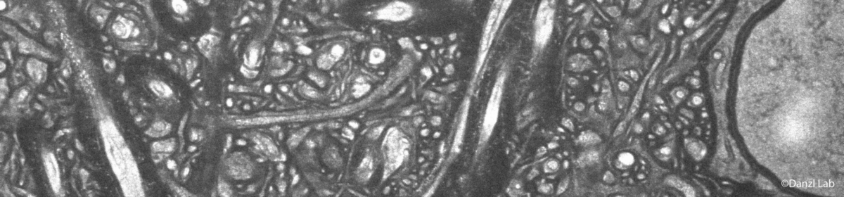
We strive to extend the possibilities of optical imaging, reaching from the organ level to the subcellular nanometer range, providing views into the tissue’s functional ultrastructure at much better resolution than the limits of classical light microscopy.
The group works toward reconstructing the brain and other cells/tissues across scales with structural and molecular detail at nanoscale resolution. We employ an integrated multi-disciplinary approach covering advanced sample preparation such as expansion microscopy, optical physics including super-resolution developments (e.g. STED and single-molecule fluorescence microscopy), and tailored computational analysis including deep-learning methods.
Recent Publications
- Light-microscopy-based connectomic reconstruction of mammalian brain tissue.
M.R. Tavakoli, J. Lyudchik, M. Januszewski, V. Vistunou, N. Agudelo Dueas, J. Vorlaufer, C. Sommer, C. Kreuzinger, B. Oliveira, A. Cenameri, G. Novarino, V. Jain, J.G. Danzl.
Nature (May 2025) DOI: 10.1038/s41586-025-08985-1 -
Image-based 3D active sample stabilization on the nanometer scale for optical microscopy.
- Human hippocampal CA3 uses specific functional connectivity rules for efficient associative memory.
J. F. Watson, V. Vargas-Barroso, R. J. Morse-Mora, A. Navas-Olive, M.R. Tavakoli, J.G. Danzl., M. Tomschik, K. Rössler, P. Jonas.
Cell (Dec 2024) DOI: 10.1016/j.cell.2024.11.022 - Super-resolution expansion microscopy in plant roots.
M. Gallei, S. Truckenbrodt, C. Kreuzinger, S. Inumella, V. Vistunou, C. Sommer, M.R. Tavakoli, N. Agudelo-Duenas, J. Vorlaufer, W. Jahr, M. Randuch, A. Johnson, E. Benkova, J. Friml, J.G. Danzl.
The Plant Cell (2025) https://doi.org/10.1093/plcell/koaf006 - Imaging brain tissue architecture across millimeter to nanometer scales.
J. Michalska, , , , , C., N. Amberg, , , , R. Höftberger, S. , G., P. , J.G.
Nature Biotechnology (2023) https://www.nature.com/articles/s41587-023-01911-8 (open access). See associated News & Views article: https://www.nature.com/articles/s41587-023-02036-8. -
Dense 4D nanoscale reconstruction of living brain tissue.
P. Velicky, E. Miguel, J. M. Michalska, J. Lyudchik, D.Wei, Z. Lin, J. F. Watson, J. Troidl, J. Beyer, Y. Ben-Simon, C. Sommer, W. Jahr, A. Cenameri, J. Broichhagen, S. G. N. Grant, P. Jonas, G. Novarino, H. Pfister, B. Bickel and J. G. Danzl.
Nature Methods (2023) https://www.nature.com/articles/s41592-023-01936-6 (open access). See associated Research Briefing: http://dx.doi.org/10.1038/s41592-023-01937-5. See also: https://www.nature.com/articles/s41592-023-02158-6. - Chiral and nematic phases of flexible active filaments.
Z. Dunajova, B. P. Mateu, P. Radler, K. Lim, D. Brandis, P. Velicky, J. G. Danzl, R. W. Wong, J. Elgeti, E. Hannezo & M. Loose.
Nature Physics (2023). https://doi.org/10.1038/s41567-023-02218-w